Sacroiliac Joint Injection.
Fact Sheets | Interventional Radiology | Back & Spinal Pain Injections
Sacroiliac Joint Injection.
Introduction
Sacroiliac joints are at the lower part of the spine, where the sacral spine segments (the part of the spine below the lumbar spine) connect with the pelvic bones flanking the sacrum, known as the iliac bones. The joint is complex, with the lowermost part of the joint typically targeted for injection of cortisone.
Sacroiliac joint pain is often vague and variable, such that it may be difficult to determine whether these joints are causing a patient’s pain or not. As clinical tests for sacroiliac joint pain may be unreliable, frequently a trial injection is performed to see if the pain is alleviated. Other treatment options following a response to a cortisone injection include destroying the nerves which supply the joint with radiofrequency ablation (RFA) as a longer lasting option.
Preparation
We also strongly recommend that you bring a responsible person to drive you home afterwards.
RISKS
most of these are minor (<1%), however can be serious (<0.1%) requiring hospital admission, intravenous antibiotics and surgery.
this is fortunately also rare and slightly common in patients with bleeding disorders and on “blood thinning” medication.
Procedure
CT Guided
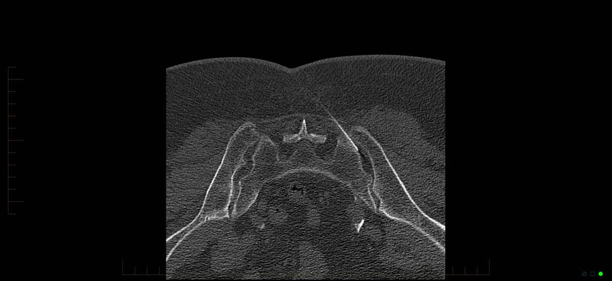
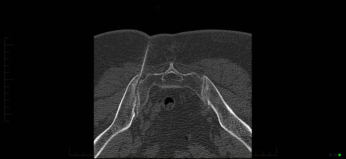
Sacroiliac Joint Injection
You will be asked to wear a gown with the selected area of the spine exposed. Sacroiliac joint injections are completed with you lying face down in a CT scanner. We will ensure that you are as comfortable as possible.
A series of planning images are performed, with the area of needle entry planned on the computer terminal and then marked on your skin. The radiologist will then clean your skin with an antiseptic wash and inject local anaesthetic into the injection site. This results in a stinging sensation which is temporary until the skin becomes numb, usually taking 10 seconds.
A fine needle is then passed through the skin and tissues, constantly manipulated under CT guidance until it enters the sacroiliac joint. A mixture of cortisone and local anaesthetic are injected which decrease inflammation and therefore pain.
Some discomfort will be felt for a short time whilst the cortisone-anaesthetic mixtures distends the joint. The local anaesthetic will then anaesthetise the joint. The effects of the treatment vary and it is difficult to predict the duration of pain relief until the procedure is performed. In some people, pain is relieved for months, others years and still for others it produces no relief at all. In the case of the latter, your doctor may recommend an alternative treatment, which may include radiofrequency ablation (RAF).
Following your procedure & recovery
At most, you will feel some minor discomfort in the back. As local anaesthetic has been injected into the spine most patients will be pain free. Patients are able to walk freely after the procedure and are observed in the clinic for 5-10 minutes. Following this, you may be discharged if you are feeling well.
You should not drive for the rest of the day. The following day you may return to work and gradually increase your activities.
Results &
Follow-Up
One of Melbourne Radiology Clinic’s specialist radiologists, a medical doctor specialising in the interpretation of medical images for the purposes of providing a diagnosis, will then review the images and provide a formal written report. If medically urgent, or you have an appointment immediately after the scan to be seen by your doctor or health care provider, Melbourne Radiology Clinic will have your results ready without delay. Otherwise, the report will be received by your doctor or health care provider within the next 24 hours.
Please ensure that you make a follow up appointment with your referring doctor or health care provider to discuss your results.
Your referring doctor or health care provider is the most appropriate person to explain to you the results of the scans and for this reason, we do not release the results directly to you.
Reminders
 Back Pain – Patient Guide
 Facet Joint Injection – Patient Fact Sheet
 Selective Nerve Root Block (SNRB)/Perineural Injection – Patient Fact Sheet
 Medial Branch Block – Patient Fact Sheet
 Epidural Injections – Patient Guide
 Discogram Injections – Patient Fact Sheet
 Sacroiliac Joint Injection – Patient Fact Sheet
 Radiofrequency Ablation (RFA) – Patient Fact Sheet
