Hamstring and Groin Injuries - Diagnostic Imaging (MRI & Ultrasound)
Muscle tears around the hip and pelvis are common, with the hamstring muscles the most frequently strained muscles.
In severe cases, hamstring injuries may be full thickness in their extent and typically require surgery, as do injuries which have extensive tendon involvement.
Groin injuries form a spectrum of injuries which may involve the adductor tendons, pubic symphysis (osteitis pubis) and also the inguinal canal, resulting in hernia formation.
Groin pain has been traditionally misunderstood, however recent advances in MRI and ultrasound, as well as improved understanding of biomechanics and anatomy of the groin has resulted in accurate diagnoses that can also gauge the severity of injury, allowing the clinician to determine whether the athlete should undergo extended rest, rehabilitation or surgery.
Case examples:
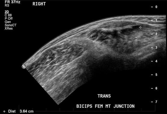
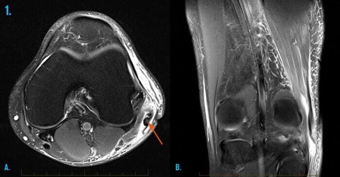
1. Hamstring injury
Axial (A) and coronal (B) MR images demonstrates a full thickness tear with retraction of the biceps femoris insertion along the outer aspect of the knee, in a young footballer. This is an unusual pattern of injury, as most hamstring ruptures occur at the tendon of origin at the level of the pelvis.
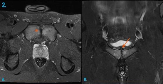
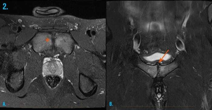
2. Osteitis pubis
Bone marrow oedema (asterisk) on both sides of the pubic symphysis is the hallmark finding of osteitis pubis. The can be associated with bone cysts and superior capsular hypertrophy (arrow). Axial (A) and coronal (B) MR images.
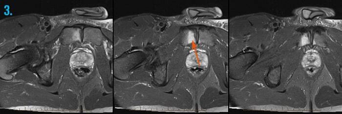
3. Osteitis pubis
MRI of the hip and pelvis demonstrates bone marrow oedema of the right side of the pubic symphysis consistent with osteitis pubis.
