
MRI Scan Ankle Injury
Twisting injuries of the ankle can often lead to a sprain of the supporting ligaments. Most sprain injuries are minor, easy to diagnose without the aid of imaging and self limiting , healing on their own accord. With ice, rest and rehabilitation, a sprained ankle will typically heal naturally; however, if the sprain is of a higher severity or if it doesn’t respond to conservative treatment, it may be necessary to undertake further evaluation, by the way of medical imaging, so as to look for the presence of tears (“sprains”) to the ligaments that support the ankle, exclude fractures and cartilage injuries, which may lead to instability. While it is possible to use ultrasound to diagnose most ligament injuries of the ankle, its inability to evaluate cartilage and joint injuries, as well as many fractures, often results in an MRI scan being undertaken in order to thoroughly evaluate and ankle injury. To rule out a fracture, an X-ray is usually typically performed prior to an MRI scan and is often suggested by the patient’s symptoms as well as the clinical findings.


How Can an MRI Scan Help?
Magnetic Resonance Imaging (MRI) uses a very strong magnetic field combined with radiofrequency waves to produce detailed images of the body. It’s safe, painless and, generally, there are no associated health risks associated with the magnetic field and radiofrequency waves. It’s become the investigation of choice for evaluating many orthopaedic and neurologic diseases and is the superior test to perform for many conditions. When using an MRI scan for ankle injury to assess for the severity of an ankle injury, magnetic fields and radiofrequency waves are used that provide high quality images of the tendons, cartilage and ligaments in the foot and ankle, that no other single imaging test is able to assess, thereby detecting stress fractures, other injuries to bone, as well as the ligaments, cartilage and tendons.
What Happens During an MRI Scan for Ankle Injuries?
At Melbourne Radiology Clinic, we have dedicated imaging equipment so as to perform an MRI scan for ankle injury.
The procedure itself is very simple and does not require any special preparation. Prior to the procedure, you’ll be asked to remove any metallic objects from your person and then lie down on the scanning table, placing your ankle in the bore of the ankle injury MRI scan machine, with your head outside of the bore.
The scan itself will take no more than 25 minutes and you will need to remain still as test is being performed. During the scan you will be provided with noise cancelling headphones and throughout the process, our technician will explain what they are doing and be in constant contact with you. Once the scan is complete, the images of your ankle will be sent to our radiologist and, as part of our routine service, we’ll provide you and your doctor with a full written report, including observations and diagnosis within 24 hours.

Book an MRI Scan for Your Ankle Injury at Melbourne Radiology Clinic
If you are suffering with an ankle injury, an ankle injury MRI scan can be booked at Melbourne Radiology Clinic either by contacting us direct by calling (03) 9667 1667 or by asking your doctor for a referral. If you have any questions you would like answering regarding our procedure, you can send an email to info@melbourneradiology.com.au or simply complete our online form. Other than MRI scan, We also offer other examinations including knee pain CT scan, X-ray for knee pain & many more healthcare imaging services in Melbourne.
Our clinic is located on the ground floor of 3-6/100 Victoria Parade in East Melbourne and our clinic hours are 08:00-19:00 Monday to Friday and 09:00-14:00 on Saturday. We look forward to seeing you.
Case examples:
1. Achilles tendon injury
MRI of the ankle demonstrates a full thickness tear of the Achilles tendon (arrow).

2. Stress fracture
MRI of the ankle shows a stress fracture (arrow) of the calcaneus (heelbone).

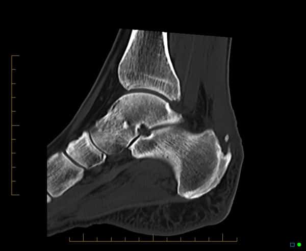
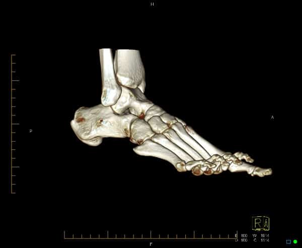
3. Use of CT to identify fractures and soft tissue calcifications
CT can be used to assess fractures of the ankle and soft tissue calcifications. 3D volume rendered images can also be obtained.
 in keeping with enthesophyte formation.")

4. Plantar fasciitis
CT can be used to assess fractures of the ankle and soft tissue calcifications. 3D volume rendered images can also be obtained.
