Hydrodilatation & Arthrography / Arthrogram.
Fact Sheets | Interventional Radiology |
Hydrodilatation & Arthrography / Arthrogram.
Introduction
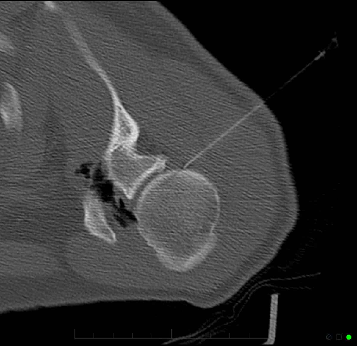
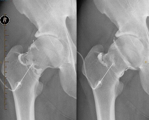
An arthrogram is a procedure usually performed under imaging guidance (using equipment such as an X-ray, CT or ultrasound machine) that involves introducing a needle into any joint of the body. It usually also involves the injection of X-ray dye (contrast) to confirm the correct depth and position of the needle.
If used solely as a pain-relieving procedure, local anaesthetic and cortisone may be injected, or other healing medications, such as glucose (prolotherapy), Autologous Blood Injection (ABI) or Platelet Rich Plasma (PRP).
An arthrogram may also be of diagnostic use. Pain that disappears following an injection of local anaesthetic into a joint usually confirms that the joint injected is the source of the patient’s pain. Though this may narrow the source of the patient’s pain, it does not determine the exact cause. For this, further imaging is usually required, such as an MRI (Magnetic Resonance Imaging) or CT (Computed Tomography) scan. If these scans are to be performed on the same day as the arthrogram, the fluid and dye injected into the joint may not only relieve the patient’s pain, but also improve the visualisation of subtle problems such as a cartilage tear.
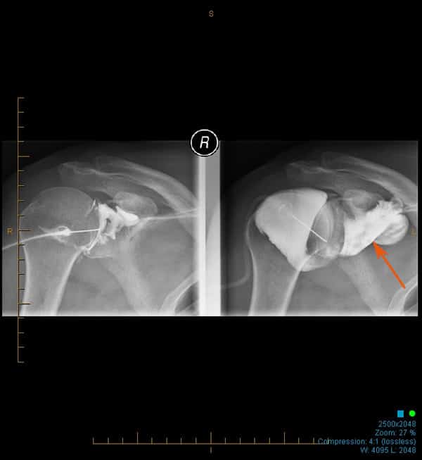
In specific circumstances, excessive inflammation in a joint may result in the lining and capsule of that joint to contract, causing pain and restriction of motion. This classically occurs in the shoulder and is commonly referred to as a ‘frozen shoulder’ (also known as adhesive capsulitis, or capsular constriction). This may be treated by the procedure of hydrodilatation, whereby an arthrogram of the shoulder is performed followed by stretching of the joint, often resulting in tearing of the capsule by injecting high volumes of saline. The effect of this is therefore twofold; pain and inflammation is reduced by the cortisone and local anaesthetic, and range of motion is improved by stretching/tearing the capsule.
Preparation
It is essential that Melbourne Radiology Clinic knows in advance of any blood thinning (or anticoagulant) medication.
These must be stopped prior to the procedure (Aspirin and Warfarin for 5 days, Plavix for 7 days & Iscover for 8 days).
We also strongly recommend to patients undergoing treatment via hydrodilatation, or undergoing an arthrogram, they attend with someone who can escort them and provide transport following the procedure.
If your diagnosis is as yet unclear, you may need to attend the clinic first for a scan, such as an ultrasound or MRI.
RISKS
most of these are minor (<1%), however can be serious (<0.1%) requiring hospital admission, intravenous antibiotics and surgery.
this is fortunately also rare and slightly common in patients with bleeding disorders and on “blood thinning” medication. Blood thinning medications that you are currently taking should be ceased (Aspirin and Warfarin for 5 days, Plavix for 7 days and Iscover for 8 days).
As these medications constantly evolve with advances in medicine, the times to cease these medications may vary with newer medications. Should you be on different medications, please contact us or the doctor who prescribed the medication for further advice.
from direct needle trauma, or as a consequence of the above mentioned complications.
Procedure
IMAGE GUIDED
Hydrodilatation & Arthrogram
After changing into a gown, you will lie on a table. The joint to be injected is then located using the relevant radiological equipment (such as an x-ray machine, ultrasound or CT scanner). A mark is then placed on your skin that correlates with the path that the needle must take to pass into the joint in a safe and reliable manner. The skin is cleansed and local anaesthetic administered. A needle is then placed into the joint at which time contrast is injected to confirm position. Occasionally, depending on varying patient body sizes, the degree of difficulty of the procedure or the condition affecting the joint, the needle may require re-manipulation.
Once correct positioning of the needle has occurred and depending on the test that has been requested, one of the three following options will take place:
For simply pain relieving injections into a joint, a small dose of cortisone and local anaesthetic will be added.
For a diagnostic arthrogram before an MRI or CT scan, a dye will be injected prior to your final and definitive scan, with or without cortisone and/or local anaesthetic as outlined in (1).
For a hydrodilatation, local anaesthetic and cortisone are first injected as outlined in (1). and then the joint stretched with fluid. In the shoulder, where this procedure is most commonly performed, a 20-40mls of fluid is added.
The arthrogram is now over. The injection site is then covered with a small dressing which may be removed later that day.
Following your procedure & recovery
Avoid any significant use of the joint that has been injected until the pain starts to resolve. This may take several days. If you hear or feel a gurgling sounds in your joint, this is normal, as air in the needle often enters the joint with the fluid and swirls about.
If you have had a hydrodilatation, we recommend that you recommence your rehabilitation programme once the discomfort from the procedure has gone, usually 2-5 days. If this has occurred sooner, you may wish to start any self directed exercises that have been prescribed in the past. Remember, as a general rule in frozen shoulder, the more movement of the joint, a better result is likely.
Results &
Follow-Up
One of Melbourne Radiology Clinic’s specialist radiologists, a medical doctor specialising in the interpretation of medical images for the purposes of providing a diagnosis, will then review the images and provide a formal written report. If medically urgent, or you have an appointment immediately after the scan to be seen by your doctor or health care provider, Melbourne Radiology Clinic will have your results ready without delay. Otherwise, the report will be received by your doctor or health care provider within the next 24 hours.
Please ensure that you make a follow up appointment with your referring doctor or health care provider to discuss your results.
Your referring doctor or health care provider is the most appropriate person to explain to you the results of the scans and for this reason, we do not release the results directly to you.


