
Foot and Ankle Injuries - Diagnostic Imaging (X-ray, MRI, CT Scan & Ultrasound)
Twisting injuries of the ankle leading to a sprain of the ligaments is one of the most frequently encountered diagnosis made on the sporting field.
Most injuries do not require imaging for a diagnosis, as most sprains are mild and the diagnosis is clear, with the athlete rehabilitating well and returning to competition promptly.
However, in certain cases, further evaluation may be required, in particular to evaluate for the presence of tears of multiple ligaments, instability, small fractures and cartilage shearing defects, which are suspected when the injury appears severe from the outset or the athlete is not progressing as well as can be expected.
An X-ray to rule out a fracture followed up by an MRI scan is the mainstay of ankle evaluation in this setting.
Ultrasound may be also used to diagnose ligament injuries, however is unable to evaluate joint and cartilage injuries. It is excellent at evaluating tendon injuries (including partial and full thickness tears) as well other soft tissue overuse disorders of the foot, including intermetatarsal bursitis, cysts/ganglia, Morton’s neuroma and plantar plate tears.


Case examples:
1. Achilles tendon injury
MRI of the ankle demonstrates a full thickness tear of the Achilles tendon (arrow).

2. Stress fracture
MRI of the ankle shows a stress fracture (arrow) of the calcaneus (heelbone).

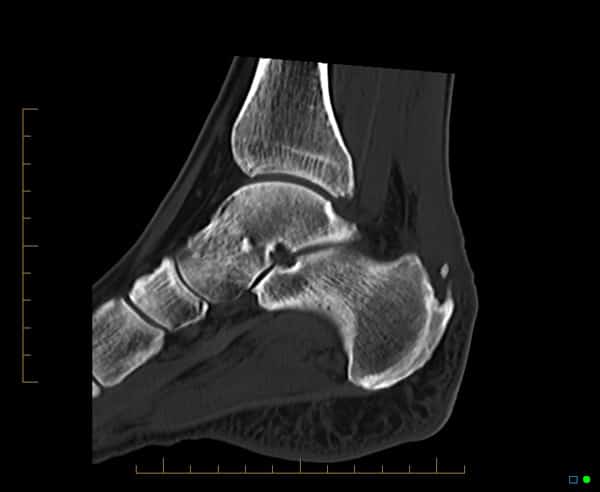
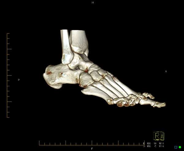
3. Use of CT to identify fractures and soft tissue calcifications
CT can be used to assess fractures of the ankle and soft tissue calcifications. 3D volume rendered images can also be obtained.
 in keeping with enthesophyte formation.")

4. Plantar fasciitis
CT can be used to assess fractures of the ankle and soft tissue calcifications. 3D volume rendered images can also be obtained.
