
Knee Injuries - Diagnostic Imaging (MRI & CT Scan)
The importance of knee mobility and balance cannot be overestimated in the athlete who requires to maneouvre rapidly between opponents, pivoting and cutting across the sporting field. All too frequently, a seemingly innocuous fall or twisting injury may result in complete disruption of the most important stabilizer of the knee, the anterior cruciate ligament (ACL). Many ACL injuries are unfortunately associated with other injuries, such as tears of the cartilage lining the joint, as well as special stabilising flat cartilaginous structures known as menisci and other ligament tears. Very rarely, all ligaments may be torn, leading to dislocation.
Degeneration of tendons (tendinosis) in front of the knee joint, also known as ‘jumper’s knee’ involves the patellar tendon and occurs in association with patellar maltracking, quadriceps tendinosis and early arthritis of the patellofemoral joint. The patellar tendon may be injected with blood (autologous blood injection and platelet rich plasma), sclerosant injection or the patient’s own cells which have been laboratory amplified (Autologous Tenocyte Implantation).
Ultrasound is a sensitive modality used to diagnose many soft tissue disorders of the knee as well as guide a needle both safely and accurately.
CT can be used to diagnose underlying patellar maltracking by measuring the shape, relations and position of the patella (kneecap).
Both CT and ultrasound can be used to perform a biopsy, where a small fragment of tissue is removed for the purposes of a diagnosis, importantly to exclude the diagnosis of a tumour.


Case examples:
1. Anterior cruciate ligament (ACL) tear
MRI of the knee demonstrates a full thickness tear of the anterior cruciate ligament (ACL). The ligament is typically taught, unlike this case (arrows).

2. Meniscus injury
MRI of the knee confirms the presence of a large tear of the lateral meniscus (arrow). Compare the appearance of the torn meniscus with the normal medial meniscus (circled) which is triangular and hypointense (dark).

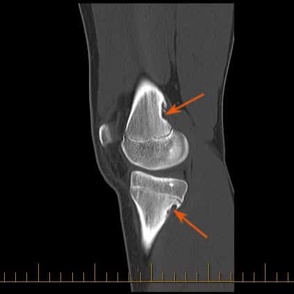
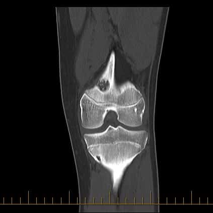
3. Use of CT to identify bone lesions
CT can be used to assess focal bone lesions, such as in this case where two cortical desmoids (arrows) are noted. Cortical desmoids are an incidental finding and require no further follow up.


