Sports Imaging
Elbow Injuries - Diagnostic Imaging (MRI)
The most common radiological diagnosis when interpreting scans of the elbow is probably “tennis elbow” or degeneration of the tendon on the outer side of the elbow known as the common extensor origin. This tendon gives rise to most of the muscle that form tendons at the level of the wrist and are involved in finger extension.
Tennis elbow is common in athletes from all sporting backgrounds. Degeneration of the tendon on the inner side of the elbow – the common flexor tendon, is known as golfer’s elbow.
Numerous treatment options exist, the most popular being Autologous Blood Injection (ABI) and Platelet Rich Plasma (PRP). More recently, Autologous Tenocyte Injection (ATI), has become available where the patient’s tendon cells are harvested, artificially grown and then reinjected. ATI is well tolerated and a safe procedure.
Case examples:
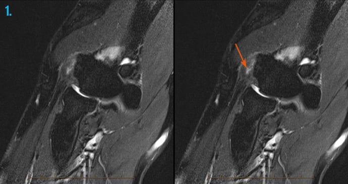
1. Tennis elbow
MRI of the elbow in an athlete with “tennis elbow” demonstrates a tear (arrow) of the common extensor origin tendon, diagnostic of this condition.

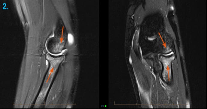
2. Bone bruising
MRI of the elbow in a young girl following a fall on an outstretched hand shows bone marrow oedema of the capitellum (upper arrow) and the radial head, neck and shaft (lower arrow) consistent with bone bruising.

