

Diagnostic Imaging for Hamstring Injury.
Radiation-Free Hamstring Tear Ultrasound.
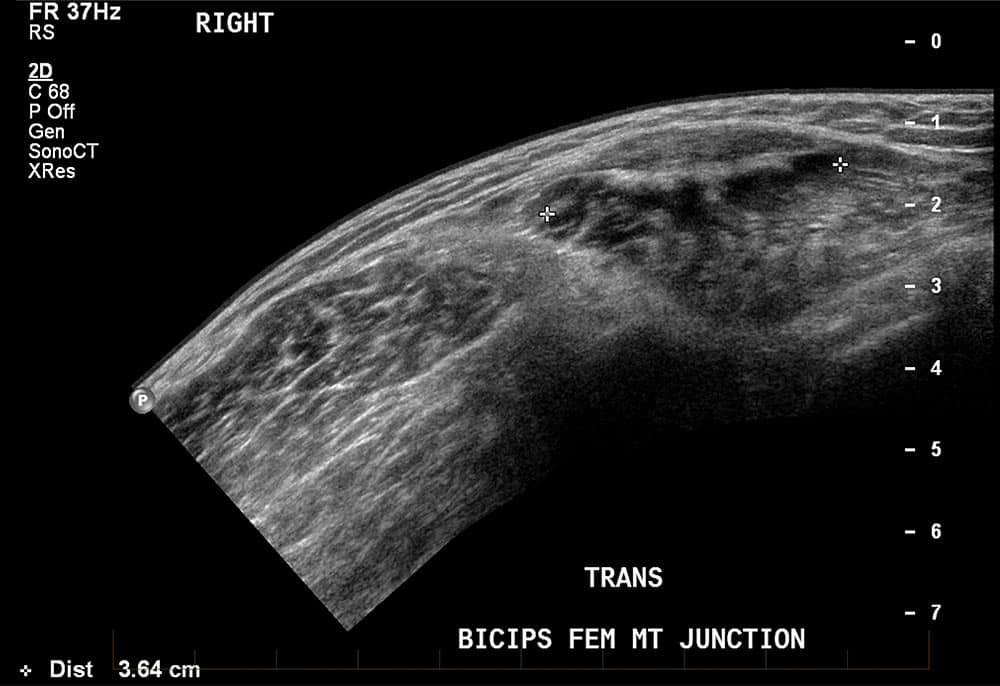
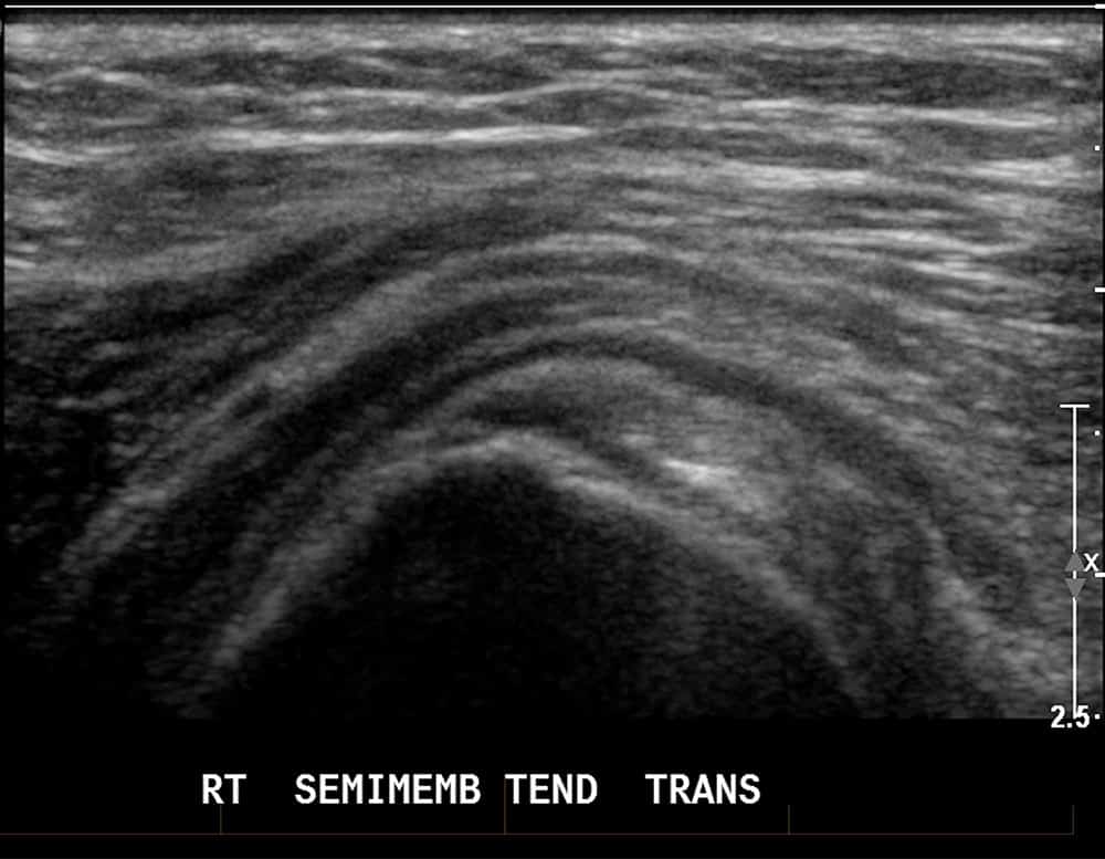
A hamstring strain or tear can occur while engaging in any type of sport or activity which involves sudden, explosive movements and can affect the semitendinosus, semimembranosus or biceps femoris hamstring muscles at the back of the thigh.
In many cases, diagnostic imaging will be required to obtain a detailed view of the extent of the injury. Ultrasound is a preferred method of diagnostic imaging for hamstring injury as it’s painless, free from radiation and the results are available quickly.


Rapid Hamstring Tear Injury Diagnosis.
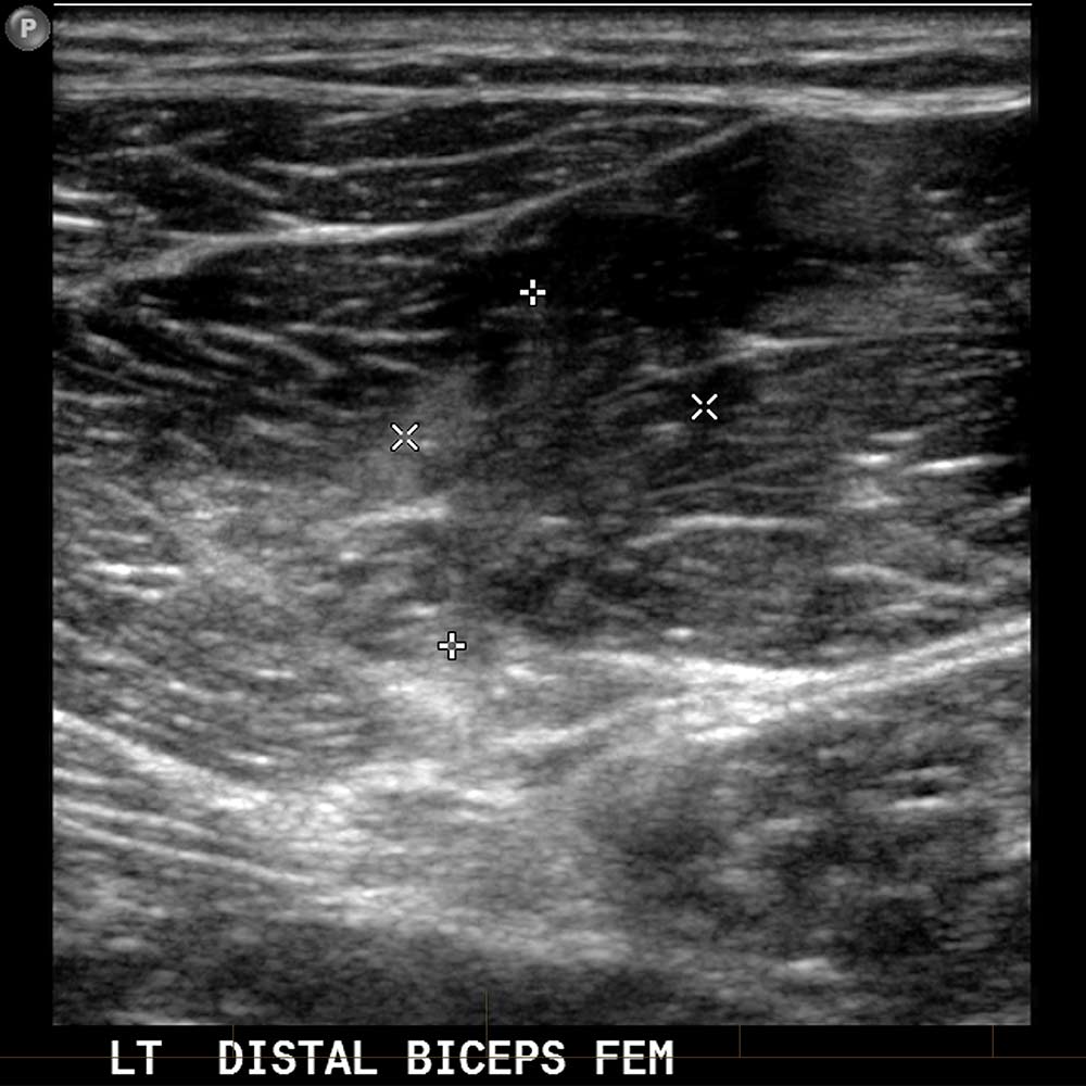
Hamstring tears can range in severity from a mild pull, to a moderate partial tear or complete tear where the tendon completely detaches from the pelvis or shin. In some cases, the tear may even pull away part of the bone (known as an avulsion fracture).
Where the injury extends into deep portions of the hamstring muscles or significant residual scarring from a previous injury is present, magnetic resonance imaging (MRI) may be recommended. In the case of an avulsion fracture (where part of the bone has also detached), an X-ray examination may also be required.
Based on the findings, your doctor will recommend if surgery is required or if the injury can be managed with non-surgical physical therapy.
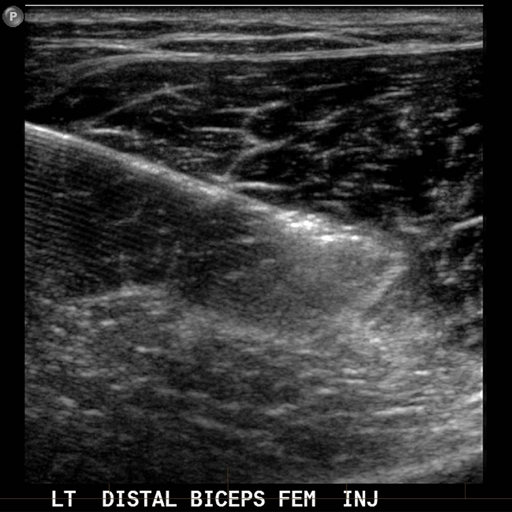
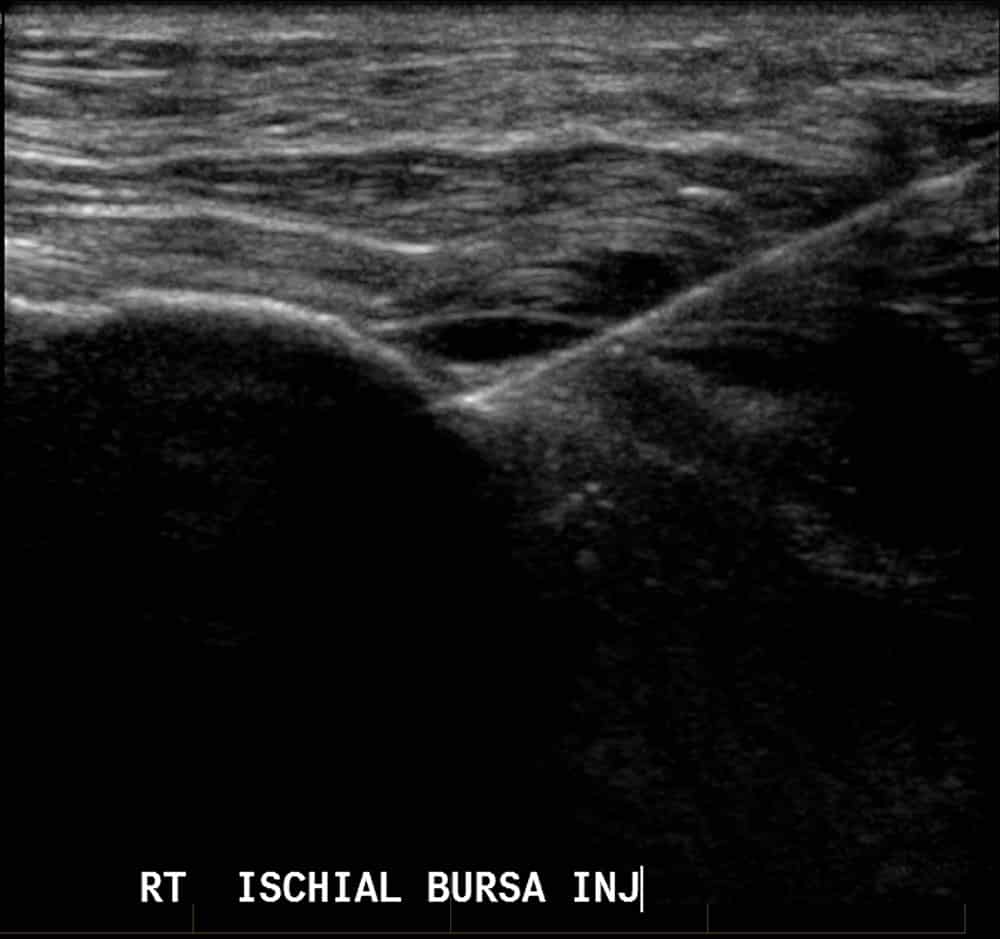
Ultrasound-Guided Hamstring Injections.
Where your doctor recommends you undergo ABI or PRP injections, the procedure can be performed with ultrasound guidance to ensure the injection is delivered precisely to the site of the injury without causing damage to surrounding tissue.

Further Information.
We welcome referring doctors to discuss the individual imaging needs of their patients with our radiologists.
Specialist Radiologists.
