
Diagnostic Hip Ultrasound Imaging.
Real-time Imaging for Rapid Diagnosis.
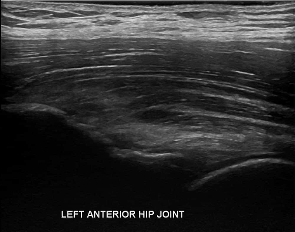
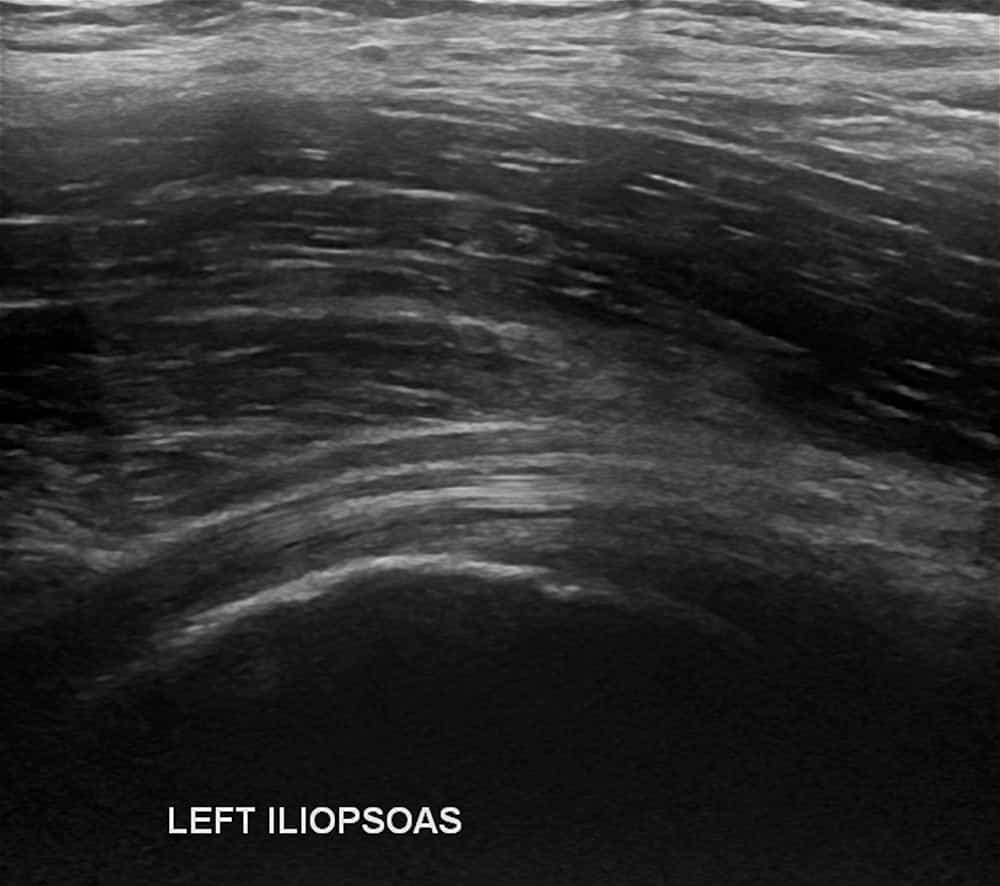
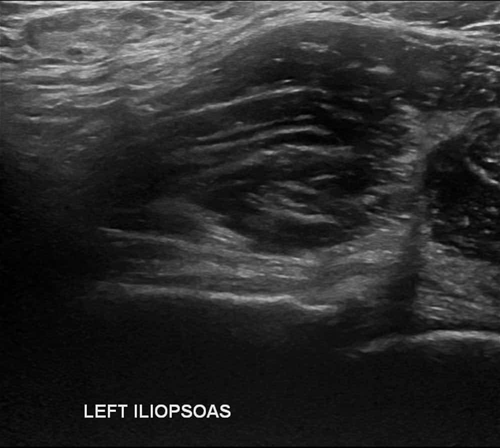
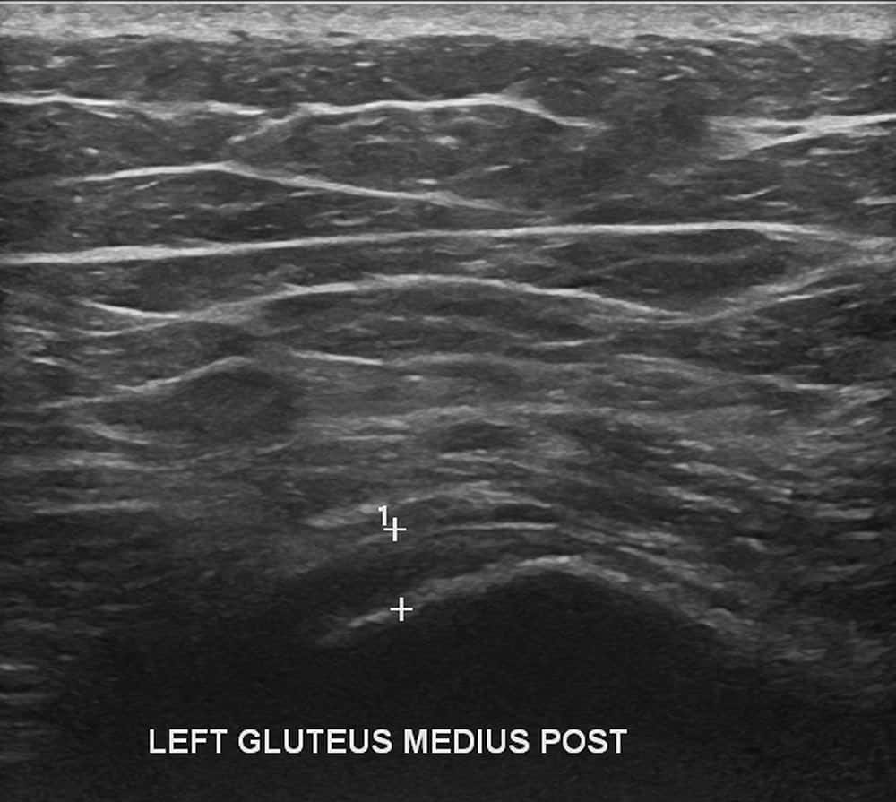
Utilising sound wave technology, hip ultrasound procedures are performed by applying a small amount of gel to the skin, before gently gliding a transducer over the area to capture real-time images of the internal structures.
The detailed imagery produced from the procedure allows your doctor to quickly detect any muscle tears, soft-tissue masses, infections, bleeding, fluid, tumours, cartilage changes or other hip injuries so they can develop an appropriate treatment plan.
While ultrasound is a highly effective method for diagnosing various soft tissue-related conditions, as it’s unable to penetrate bone, your doctor may request you undergo a CT scan or MRI if required.


Non-invasive, Safe and Painless.
A hip ultrasound is an excellent non-invasive diagnostic tool that is quick, painless and doesn’t require any specific preparation. While you may notice some light pressure when the transducer is applied to the hip area, patients generally shouldn’t experience any pain or discomfort during a hip ultrasound.
Paediatric Hip Ultrasound.
A hip ultrasound is used to assess for the presence of DDH in infants less than six months of age. As no radiation is emitted during a hip ultrasound, it can be safely performed.

Further Information.
We welcome referring doctors to discuss the imaging needs of their patients and the suitability of ultrasound based on their patient’s medical condition with our radiologists.
Specialist Radiologists.
