Ultrasound Medical Imaging.
Ultrasound technology uses inaudible soundwaves to deliver superb images without the use of radiation. Ultrasound image quality has undergone dramatic improvement over the last several years.
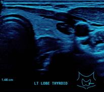
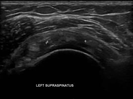
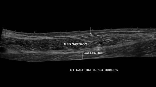
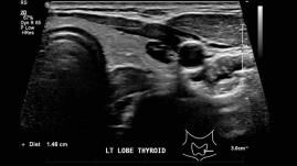
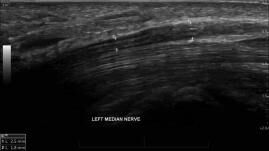
Ultrasound is of particular use in the diagnosis of soft tissue conditions and as such, it is routinely used to evaluate the solid abdominal and pelvic organs, the breast, muscles, tendons and ligaments, as well as other small body parts, such as the thyroid gland and lymph nodes.
Since images are performed in real time, ultrasound is ideal for performing procedures under imaging guidance, such as a pain relieving injection, the treatment of tendon injuries as well as taking a small specimen of tissue for analysis, known as either a Fine Needle Aspiration (FNA) or Biopsy.
Doppler Ultrasound.
(Vascular Ultrasound Imaging)
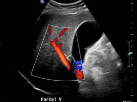
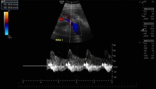
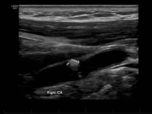
Ultrasound may also be used to visualise the arteries and veins of the limbs and the neck with the use of Doppler technology. As such, it can reliably detect narrowing of the arteries, varicose veins and blood clots.
Doppler ultrasound (also referred to as vascular diagnostic imaging) is a painless, non invasive means of measuring blood flow. For example Doppler ultrasound can detect problems with abnormal blood flow, where flow may be reduced in veins due to clot (thrombosis), known as Deep Vein Thrombosis (DVT). Ultrasound can also detected narrowing (stenosis) of arteries due to conditions such as atherosclerosis (“hardening of the arteries”); common areas include the neck (carotid arteries) as well as the arteries of both lower limbs.
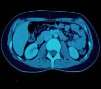
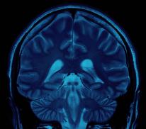
As ultrasound waves cannot penetrate bone and gas, ultrasound is not used to look inside a joint, bone or gas containing organs, such as the lungs or bowel. If these structures also require evaluation, then a CT scan or MRI may be more suitable.
Further Information.
Referring doctors are welcome to discuss with our radiologists the imaging needs of their patients and whether ultrasound is suitable for their patient’s medical condition.
Specialist Radiologists.
MSK & MRI Fellowship Trained Radiologists
