CT Scan Radiation & X-Ray Radiation Safety.
Fact Sheets | Radiation Safety
Radiation Safety.
Overview
Radiation is a type of energy that travels through space and that all living creatures require for life. For example, light is a type of radiation, as is heat. Other forms of radiation include radiowaves used in radio transmission and microwaves for cooking. Radiation differs in terms of its wavelength. The shorter the wavelength, the more damage it produces to tissue.
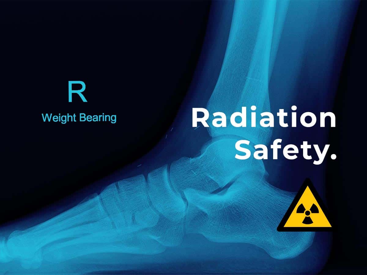
At Melbourne Radiology Clinic, only two types of scanners use radiation: a CT scanner and X-ray machine. This type of radiation is called ionising radiation and enters and leaves the body providing important diagnostic information. It can however, be harmful if excessive exposure occurs to sensitive organs.
An ultrasound and MRI (Magnetic Resonance Imaging) scanner which are available at Melbourne Radiology Clinic DO NOT use radiation.

Risks of Radiation

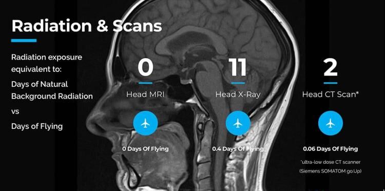
It is important to put this into context with radiation that we are exposed to every day; this is called “background radiation”. This is the radiation that we are exposed to from outer space (cosmic rays), decay of natural elements around us, as well as radon gas that collects in buildings.
The amount or dose of radiation received is expressed in units known as the millisievert, or mSv. On average, we receive 2mSv over a year from background radiation (Ref: https://www.aci.health.nsw.
A chest X-ray will result in a dose of 0.1mSv, or alternatively, 10 days equivalent of background radiation.
What does this mean and what is the risk of this dose?
The chances of dying from cancer due to a single chest X-ray is one in a million, which is also the same risk of dying from cancer from smoking ONE cigarette or drinking half a bottle of wine.
Other activities that we may engage in that have a 1 in a million risk of dying are daily activities which wouldn’t give a second thought to, such a riding a bicycle for 15km, travelling in a car for 100km or taking the contraceptive pill for 2.5 weeks.
A CT of the abdomen results in a dose of 10mSv CT scan radiation, or approximately 3 years of background radiation. A radiotherapy dose to treat cancer may easily exceed 20,000-50,000mSv.
The amount of radiation used in X-rays for medical imaging is very small and the possible benefits of the X-ray far outweigh the risks, otherwise the scan wouldn’t be ordered by your doctor. The radiation enters and then leaves your body in an instant. No radiation remains behind or lingers.
Melbourne Radiology Clinic has recently (2018) invested in a new ultra-low dose CT scanner (Siemens SOMATOM go.Up) with the level of CT Scan radiation dose reduction exceeding even our own expectations. Many CT examinations, particularly those of the peripheral skeleton, are currently at a lower dose than standard digital radiographs.
RADIATION SAFETY & YOU
It is important to be aware that all the equipment at Melbourne Radiology Clinic is routinely serviced and undergoes safety checks to ensure no radiation leaks occur. We also minimise the number of views we take to make a diagnosis and if need be, we may perform or recommend a different test that uses less or no radiation. All our staff have received the appropriate training in radiation safety and are credentialled as well as licensed to use our equipment.

Pregnant or possibly pregnant women should inform the staff at Melbourne Radiology Clinic.
Radiation of the developing fetus must be kept to a minimum, however if the mother is suffering from a life threatening condition, then the risk of NOT doing the scan may outweigh the small risk to the fetus. Other alternative tests may be used as a substitute. Wherever possible, direct exposure to the pelvis should be avoided. Radiation to other body parts does result in a smaller dose to the fetus by the process of “scatter”, however is significantly smaller than exposure to the primary X-ray beam that is, when the X-ray is directed at the pelvis.
If you are concerned about the dose you may receive in an X-ray please discuss this with your doctor or call Melbourne Radiology Clinic.
