Autologous Tenocyte Implantation & Autologous Tenocyte Therapy
Fact Sheets | Interventional Radiology | Musculoskeletal Imaging
Autologous Tenocyte Implantation (ATI)
/ Autologous Tenocyte Therapy (ATT).
Introduction
Tendons connect muscle to bone, transmitting the forces generated by muscle contraction onto bones and thus are crucial for the mobility of all joints. Like any tissue, tendons may be damaged due to trauma and also undergo age related “wear and tear” (degeneration). With increasing age, the number of tendon cells (tenocytes) decline, resulting in the tendon having limited ability to repair itself. Ultimately, the tendon loses its integrity and develops tears, consequently unable to withstand normal day to day activities. This most commonly manifests as joint related pain and loss of function. Overall, this process of tendon degeneration is known as tendinopathy (or tendinosis), commonly (though incorrectly) colloquially referred to as “tendinitis”. Unfortunately, the body’s reaction is unable to reverse this process.
ATI is however able to artificially replenish the tenocyte population, with the aim of improving tendon structure, strength and function, thereby minimising tendon related pain.
Naturally, any tendon of the body can be injected, with the most commonly injected areas being the elbow (common extensor tendon), knee (patellar tendon), ankle (Achilles tendon), hip/buttock (gluteal and hamstring tendons) as well as the shoulder (supraspinatus tendon).
Preparation
It is essential that Melbourne Radiology Clinic knows in advance of any blood thinning (or anticoagulant) medication.
These must be stopped prior to the procedure (Aspirin and Warfarin for 5 days, Plavix for 7 days & Iscover for 8 days).
We also strongly recommend that you bring a responsible person to drive you home afterwards.
***
Important information to tell your doctor prior to treatmentFortunately serious side effects are rare, however if you have an existing condition, this must be discussed with your referring doctor before having this treatment. People with local skin or systemic infections are at greater risk of having an infection spread surrounding soft tissues and joints. Therefore, if you have a skin infection, which may include wounds, boils or rashes, please tell your doctor or arrange to have the procedure performed at a later date.
RISKS
most of these are minor (<1%), however can be serious (<0.1%) requiring hospital admission, intravenous antibiotics and surgery.
this is fortunately also rare and slightly common in patients with bleeding disorders and on “blood thinning” medication. Blood thinning medications that you are currently taking should be ceased (Aspirin and Warfarin for 5 days, Plavix for 7 days and Iscover for 8 days).
As these medications constantly evolve with advances in medicine, the times to cease these medications may vary with newer medications. Should you be on different medications, please contact us or the doctor who prescribed the medication for further advice.
from direct needle trauma, or as a consequence of the above mentioned complications.
As the cells are derived from your body, there is no risk of developing side effects, rejection or infectious disease transmission.
Procedure
Ultrasound Guided
ATI/ATT Injections
ATI is a two stage process. Both stages are simple and quick, taking less than 30 minutes each and are well tolerated.
Stage 1
Harvesting of Tendon Cells
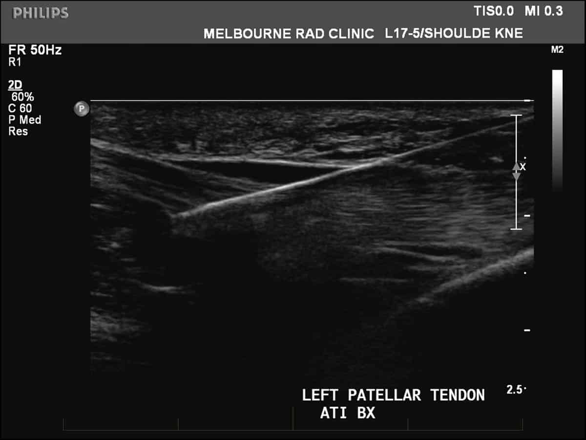
You will be asked to wear a gown, lie down on a couch on your back with the selected area of the body exposed. We will ensure that you are as comfortable as possible. Initially a small specimen of cells is harvested from a normal tendon. The patellar tendon (below the patella, or knee cap) is typically used as it easily accessible and of adequate size. An ultrasound examination takes place first in order to confirm that the tendon is normal to guarantee the quality of the specimen. Once this is confirmed, the radiologist will clean your skin with an antiseptic wash and inject local anaesthetic into the skin and soft tissues. This results in a stinging sensation which is temporary until the skin and soft tissues becomes numb, usually taking 10-30 seconds. Under ultrasound guidance, a biopsy needle is used to harvest two small core specimens of the patellar tendon.
The specimens are then placed in a culture medium and grown over a period of several weeks, usually 4- 6 weeks, at the Orthocell biotechnology laboratory in Perth. This process typically produces five million healthy tendon cells (tenocytes). Blood will also be drawn from your arm for the purposes of growing your cells in your serum (the fluid component of your blood). You will then be contacted with respect to the timing of the second injection, at which point, the cells are delivered to Melbourne Radiology Clinic.
Stage 2
Injection of tenocytes into painful tendon
The painful tendon to be injected is identified, once again using an ultrasound scanner. The skin is cleansed and anaesthetised as for stage 1. Under ultrasound guidance, the tenocytes are injected safely into painful tendon and into the area of maximal abnormality.
Following your procedure & recovery
At most, you will feel some discomfort in the injected areas. As local anaesthetic has been injected, you will be pain free for several hours. You will be able to walk freely after the procedure and discharged at your leisure. If you do experience pain once the local anaesthetic wears off, simple measures, such as rest for 48 hours, application of a cool compress, taking regular paracetamol and avoiding alcohol, are usually all that are required.
After the initial 48 hours of rest, you may commence gentle stretching for the first two weeks.
A rehabilitation programme focusing in eccentric exercises under the supervision of a health professional such as physiotherapist or sports doctor is commenced. A separate fact sheet from Orthocell will be provided to you at the time of the stage 2 procedure.
The effects of ATI take several weeks to months to be noticed, which reflects the time taken for the tendon population to regenerate and produce functional and healthy tendon tissue.
Results &
Follow-Up
One of Melbourne Radiology Clinic’s specialist radiologists, a medical doctor specialising in the interpretation of medical images for the purposes of providing a diagnosis, will then review the images and provide a formal written report. If medically urgent, or you have an appointment immediately after the scan to be seen by your doctor or health care provider, Melbourne Radiology Clinic will have your results ready without delay. Otherwise, the report will be received by your doctor or health care provider within the next 24 hours.
Please ensure that you make a follow up appointment with your referring doctor or health care provider to discuss your results.
Your referring doctor or health care provider is the most appropriate person to explain to you the results of the scans and for this reason, we do not release the results directly to you.

