Hip Injuries - Diagnostic Imaging (CT Scan, MRI, Ultrasound)
Hip problems are increasingly becoming frequently recognised as a common source of pain in the athlete. Increasing demands on the lower spine and pelvis have created ever increasing demands on the hip, which must simultaneously provide strength, speed, balance and stability. Not surprisingly, hip disorders can adversely affect an athlete's career.
As the hip is a ball and socket joint, formed by the upper thigh bone (femur) and pelvis (acetabulum), incongruity of one or both bones may result in abutment (impingement), causing cartilage wear and tear (degeneration), ultimately premature osteoarthritis. Not surprisingly, this condition is known as femoroacetabular impingement, or ‘FAI‘. As FAI may result in decreased hip range of motion, increased stress is places on the pelvis, thus predisposing the pubic symphysis also to degeneration, commonly referred to as osteitis pubis (OP).
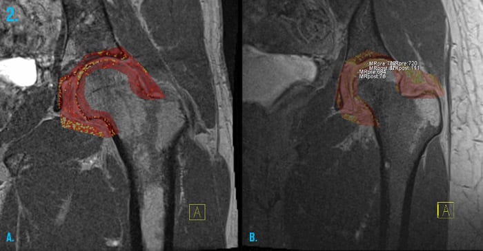
Early identification of cartilage wear and tear may be made by a specific MRI scan known as delayed Gadolinium Enhanced MRI evaluation of Cartilage (dGEMRIC) and is an important tool in evaluating which patients may benefit from reconstructive surgery. Hip pain injections may be administered as part of a treatment.
Case examples:
1. Femoroacetabular impingement (FAI) - CT guided hip injection
CT guided hip injection of cortisone for femoroacetabular impingement (FAI), a condition where the femoral head is not completely spherical (arrow) and results in abutment against the acetabulum (asterisk)

2. dGEMRIC Hip MRI
Delayed Gadolinium Enhanced MRI evaluation of Cartilage (dGEMRIC) in a young patient shows early stages of cartilage degeneration that is irreversible (white and yellow color pixels; normal cartilage = red pixels).

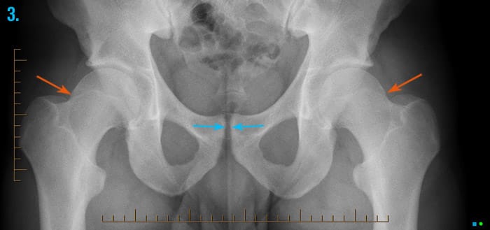
3. Athletic chronic groin injury (Cam type FAI with osteitis pubis)
Bilateral prominence of the femoral heads along the lateral (outer) aspect (orange arrows), is consistent with cam type femoroacetabular impingement. This may result in decreased mobility of the hip, placing increased stress on the pelvis, particularly the pubic symphysis, predisposing the athlete to the development of osteitis pubis (blue arrows).[1]

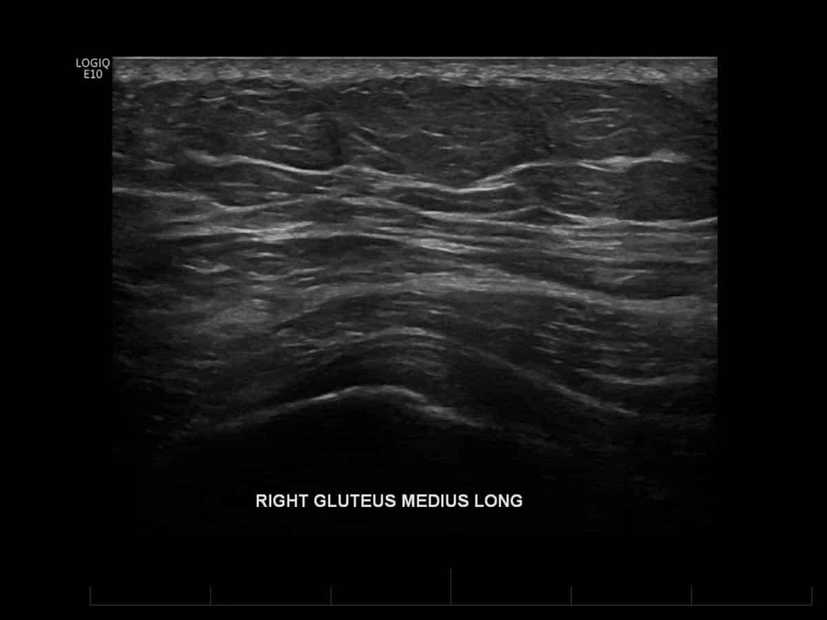
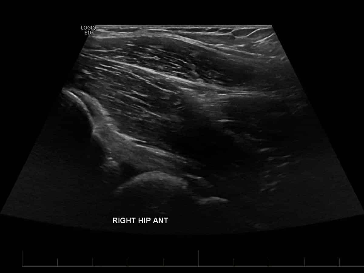
4. Hip Ultrasound
Ultrasound of the hip is used predominantly to assess for the soft tissues (“extra-articular”) structures that lie external to the hip joint, namely the muscles, tendons, bursae and nerves. Assessment of the insertion of the gluteal tendons for the presence of tendinosis, tears and/or bursitis would be the most common reason to perform an ultrasound of the hip, in which case, a cortisone injection into the trochanteric bursa may performed in the same sitting.
Other tendons that can be assessed include the hamstring origin from the ischial tuberosity (posterior hip) as well as the iliopsoas tendon and bursa (anterior hip). Hernias of the groin can also be assessed, as well as the femoral and lateral femoral cutaneous nerves. Inflammation/irritation of the latter nerve is known as meralgia paraesthetica and can be treated with an ultrasound guided cortisone injection.
References:
- Verrall GM, Slavotinek JP, Barnes PG, Esterman A , Oakeshott RD, Spriggins AJ. Hip joint range of motion restriction precedes athletic chronic groin injury. J Sci Med Sport 10(6):463-6, 2007


