Melbourne Radiology Clinic specialises in the treatment of musculoskeletal pain syndromes and offers a broad range of tests to assist in obtaining an accurate diagnosis.
Ultrasound and CT guided injections
Melbourne Radiology Clinic can also use its modern imaging equipment to accurately guide a needle to deliver pain relieving medication precisely into the region that is the patient’s source of pain, with minimal trauma to surrounding tissue.
As such, these injections are useful in patients who have not responded adequately to rest, rehabilitation and medications. It is also of use in patients as a last resort prior to contemplating, or awaiting surgery.
Musculoskeletal conditions treated on a daily basis at Melbourne Radiology include:
- Bursitis - The following are an example of the many bursae of the body which which routinely require injection: subacromial (shoulder), trochanteric (lateral hip), iliopsoas (anterior hip), ischial (buttock), intermetatarsal (forefoot), prepatellar and infrapatellar (knee), retrocalcaneal and retroachilles (Achilles tendon)
- Morton's neuroma injections
- Tenosynovitis/paratenonitis, deQuervain's (wrist), Achilles tendon paratenonitis
- Ganglion/cyst aspiration: such as ganglia arising from any joint, commonly around the wrist and cysts of the knee (Baker's or popliteal cyst)
- Dupuytren's Contracture
- Plantar fasciitis
- Arthritis - Hyaluronan Injections
- Direct tendon injections - ABI, PRP & Sclerosant Injection

Bursitis: including subacromial and trochanteric bursitis
A bursa is a fluid filled sac that surrounds joints and tendons and acts like a lubricant, allowing tendons and muscles to move bones with minimal friction. Wear and tear with excessive use or increasing age may damage a bursa, causing it to become inflamed and losing its lubricating ability. This then results in pain with certain movements.
Most cases respond well to medication and rehabilitation, along with restriction or cessation of the activity that produced the symptoms in the first place.
Occasionally, symptoms may persist despite best efforts and a cortisone injection directly into the bursa is a frequently successful procedure. This is usually performed under ultrasound guidance, with the needle tip placed into the bursa under direct vision, guaranteeing precise delivery of the medication into the area where it is needed most.
Morton's neuroma injections.
Forefoot pain has many causes which may be evaluated with both ultrasound and MRI.
Common causes of forefoot pain include joint inflammation (arthritis, capsulitis & synovitis), plantar plate tears, tendinosis (“tendinitis”), bursitis and Morton’s neuroma.
A Morton’s neuroma occurs when scar tissue builds upon a nerve in between the toes known as the interdigital nerve (nerve between the digits, or toes). This is most frequently seen in women and is and is attributed to high heeled shoes. The pain is often severe and has an electric shock character to it. A non surgical option destroys the nerve by heating it using a procedure called radiofrequency ablation and usually results in relief with the one procedure.
Another form of injection based treatment is to disrupt the nerve by using highly concentrated alcohol injected under ultrasound control. This is performed weekly until symptoms improve.
References:
- Gregg JM, Schneider T, Marks P. MR imaging and ultrasound of metatarsalgia–the lesser metatarsals. Radiol Clin North Am46(6):1061-78, vi-vii, 2008
- Masala S, Fanucci E, Ronconi P, Sodani G, Taormina P, Romagnoli A, Simonetti G.Treatment of intermetatarsal neuromas with alcohol injection under US guidance.Radiol Med102(5-6):370-3, 2001
- Hughes RJ, Ali K, Jones H, Kendall S, Connell DA. Treatment of Morton’s neuroma with alcohol injection under sonographic guidance: follow-up of 101 cases. AJR Am J Roentgenol 188(6):1535-9, 2007
- Finney W, Wiener SN, Catanzariti F. Treatment of Morton’s neuroma using percutaneous electrocoagulation.J Am Podiatr Med Assoc. 1989 Dec;79(12):615-8
- Genon MP, Chin TY, Bedi HS, Blackney MC. Radio-frequency ablation for the treatment of Morton’s neuroma.ANZ J Surg.80(9):583-5, 2010


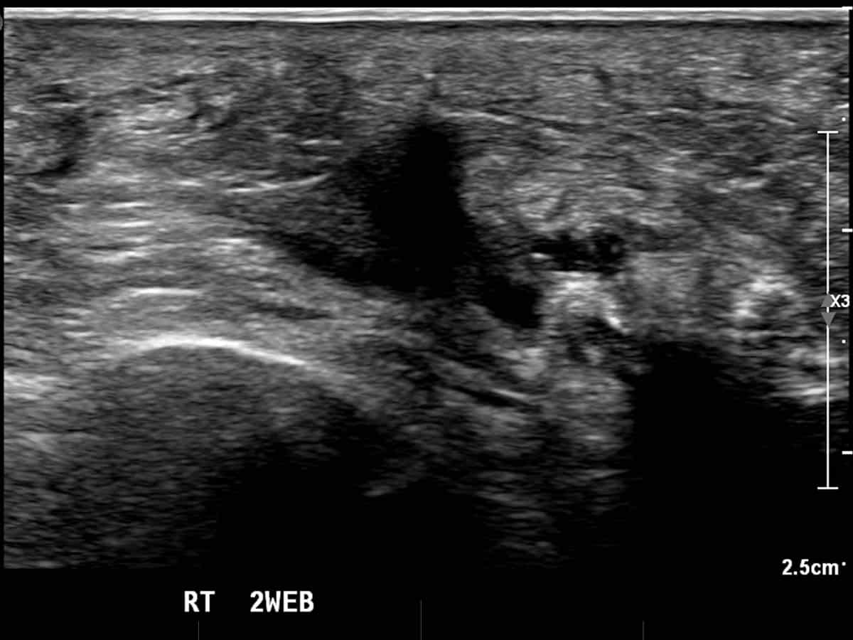
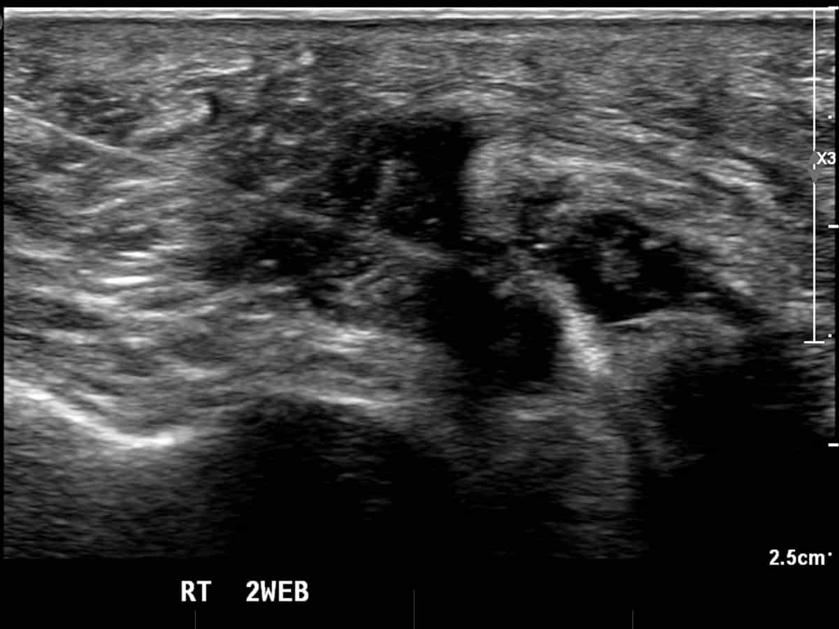
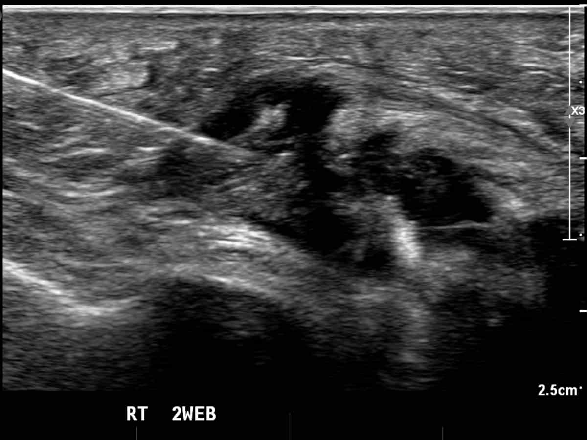
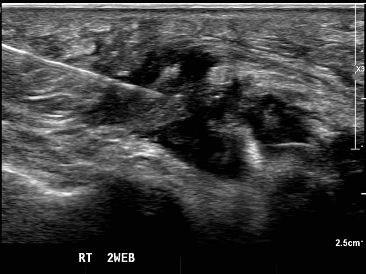
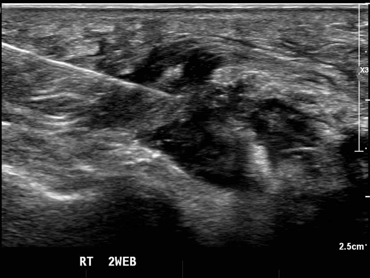
Ultrasound guided cortisone injection into a second webspace Morton’s neuroma, right side.

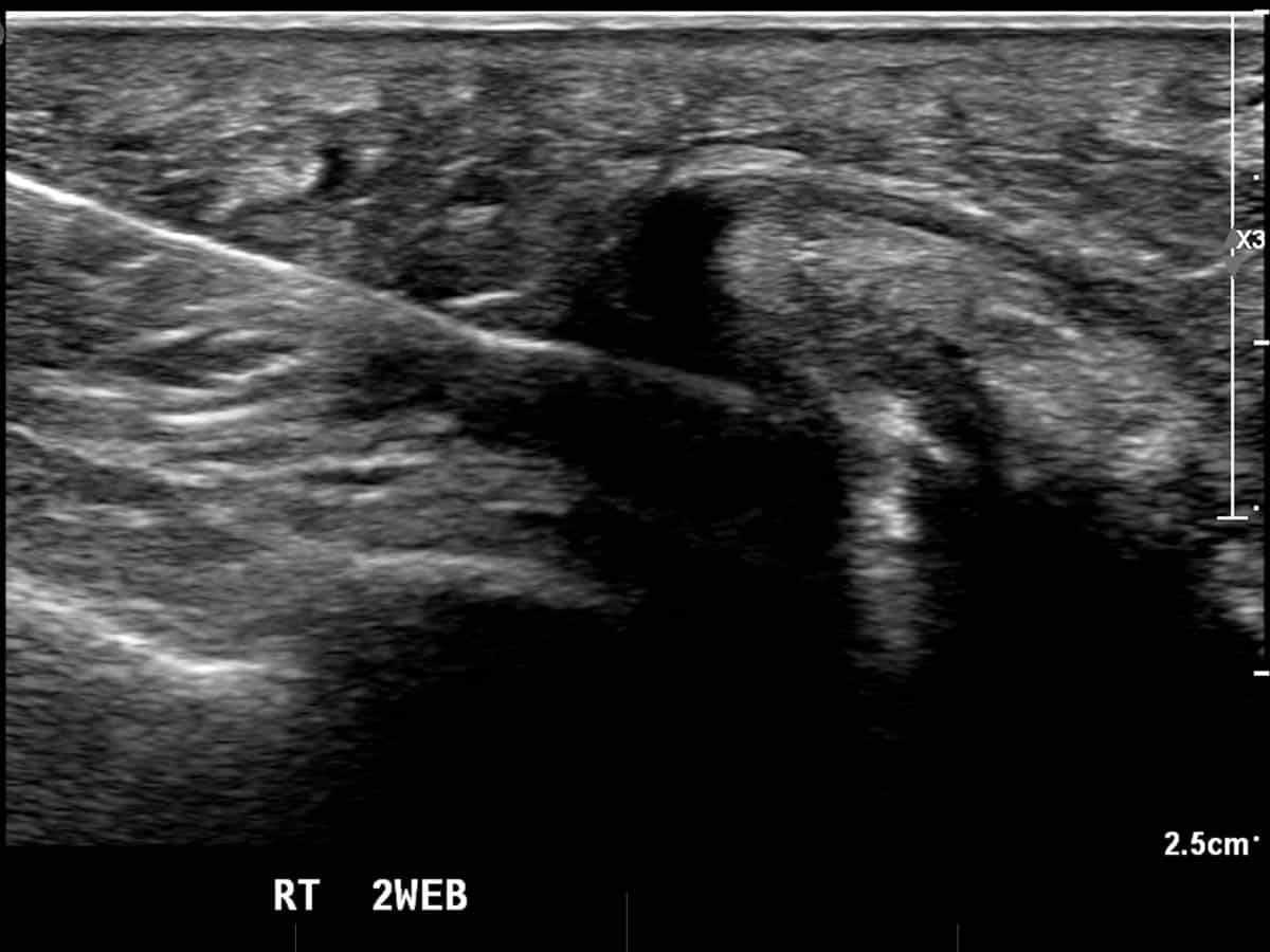
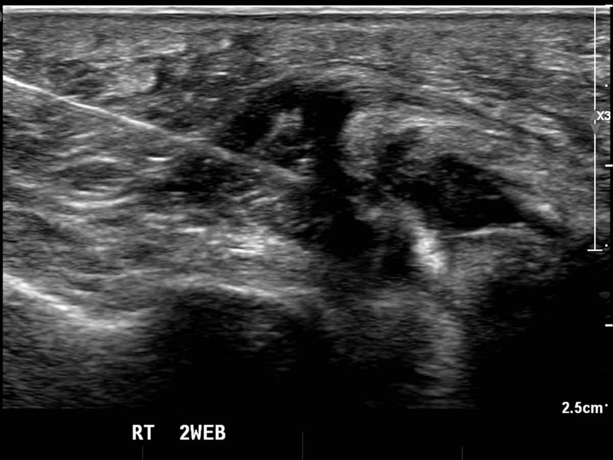
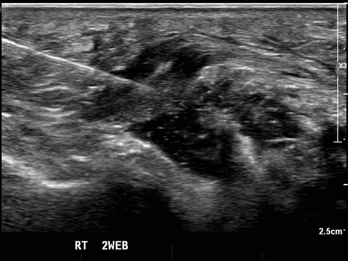
Ultrasound guided radiofrequency ablation into a second webspace Morton’s neuroma, right side.

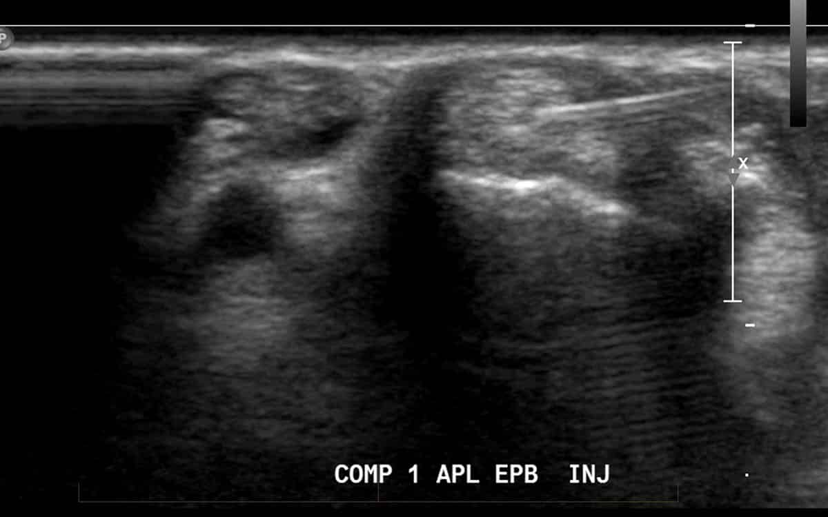
Ultrasound guided injection to treat de Quervain’s tenosynovitis
De Quervain's tenosynovitis
Repetitive manual labour may result in increased friction between tendons of the wrist. Tendons are lined by a thin membrane (tendon sheath, or tenosynovium) which when irritated by repetitive motion becomes inflamed and produces fluid. This is called tenosynovitis and may occur in any tendon of the body which possesses this lining of tissue. When it occurs in the wrist, it is known as de Quervain’s tenosynovitis, named after the person who first described the condition.
Ultrasound can be used to deliver a dose of cortisone which is in many cases curative, provided that the patient also refrains from the offending activity. This condition is common in parents who look after young children, as well as those performing previously unaccustomed exercise.
Ganglion/synovial cyst drainage, including Baker's cysts of the knee.
Joints naturally produce fluid which acts as a natural lubricant. If this fluid production is excessive, such as seen in arthritis, or following excessive use, the fluid may protrude through a weakness in the joint capsule. When this occurs, it is known as a ganglion (or synovial cyst). Often it may simply be noticed as a firm lump. Occasionally these may be painful, or compress an important nearby structure, such as a nerve.
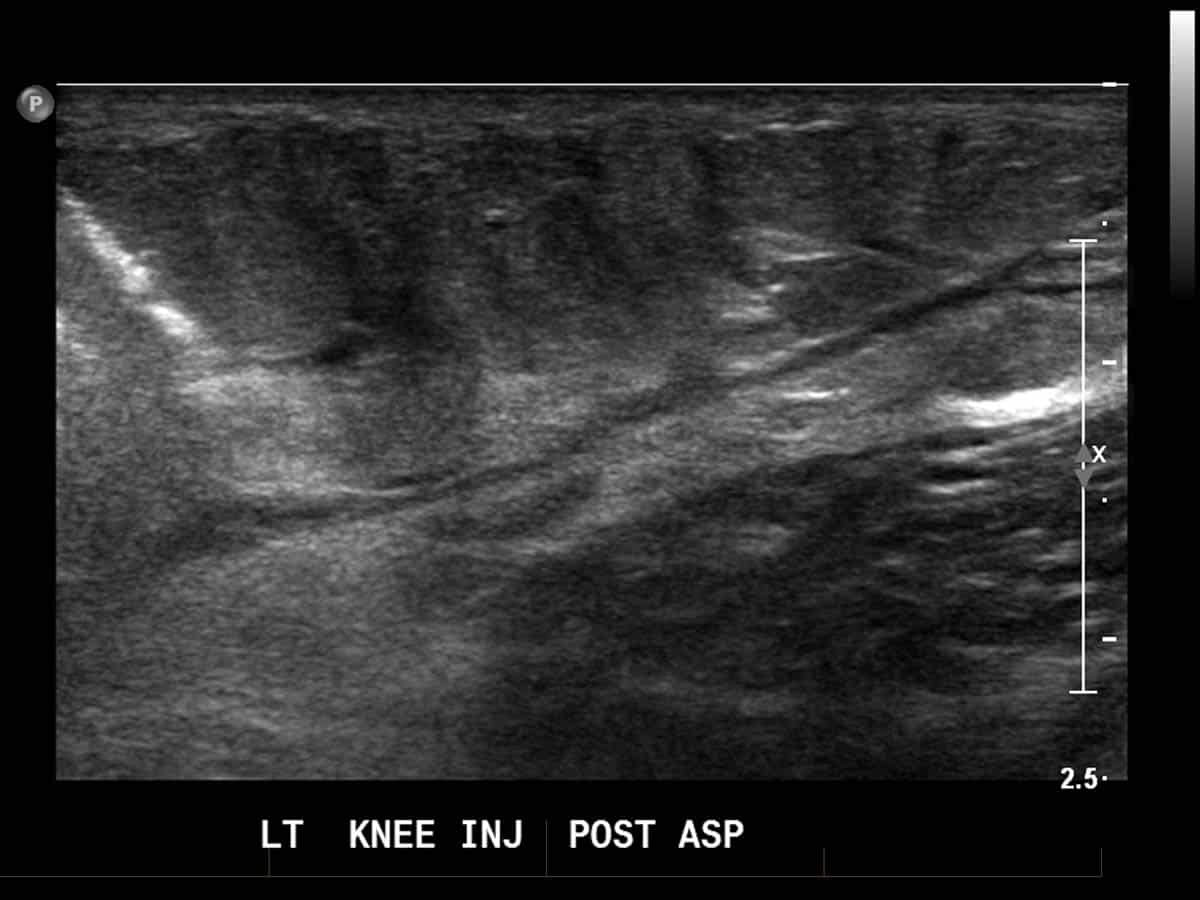
Under local anaesthetic and using ultrasound guidance, the fluid can be removed and possibly avoid the need for surgery. This is a useful technique in conditions where the cyst is in a part of the body that is difficult to operate on, and as such, treatment options are limited. This is most commonly seen in a Baker’s cyst, which occurs behind the knee.


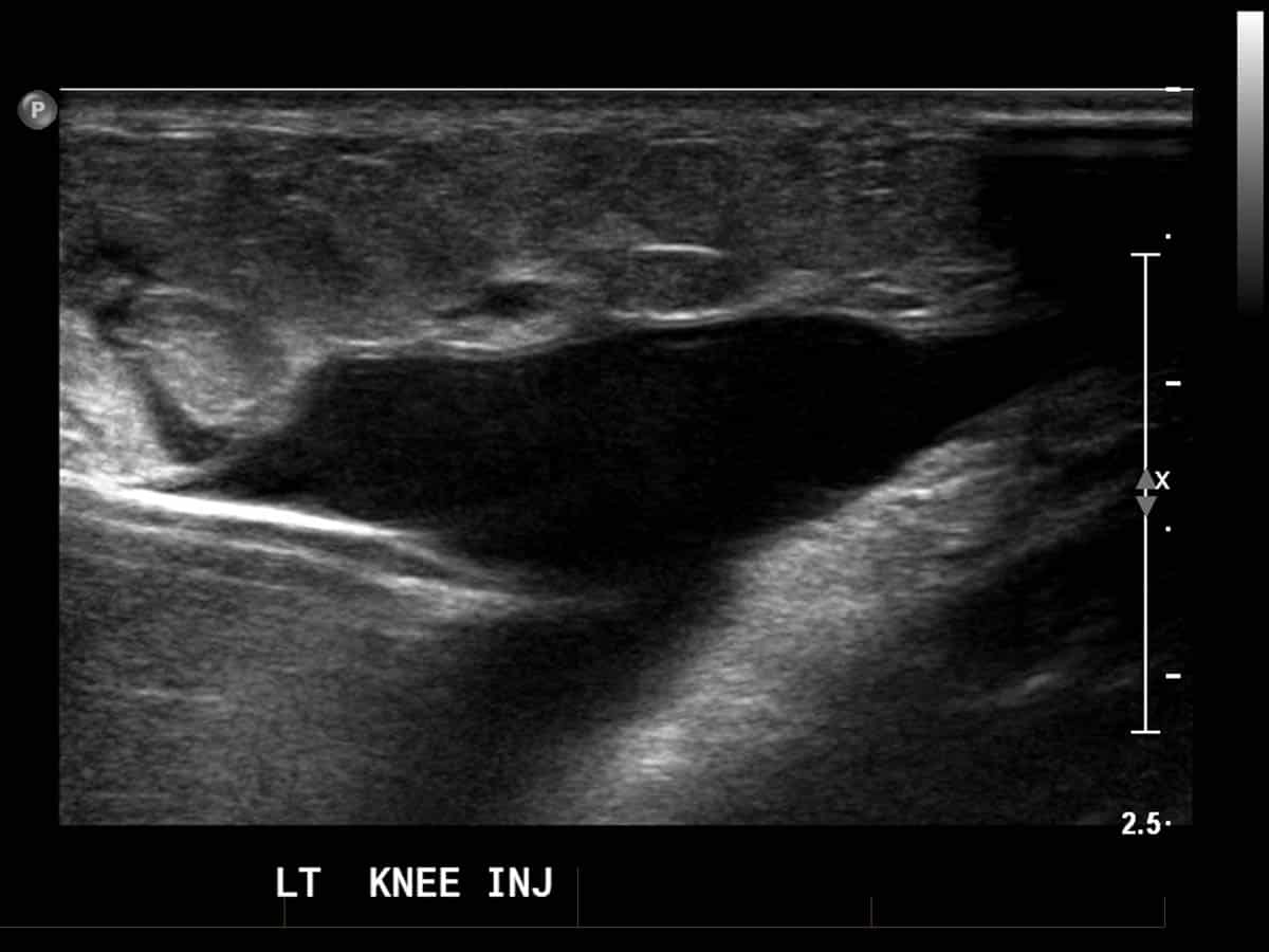
Ultrasound guided drainage (aspiration) of a superficial collection of fluid around the knee joint following trauma. (Pre and Post aspiration)
Dupuytren's contracture.
In certain individuals, a build up of scar-like tissue, similar to that occurring in patients with a frozen shoulder, occurs in the palm of the hand, resulting in a firm and often painful nodule under the skin. If this progresses, contraction of the adjacent tissues occurs, including the tendons to the finger, which may make straightening the finger impossible. Ultrasound can be used to accurately deliver a dose of cortisone into the scar tissue with the aim of resolving symptoms. Best results are achieved when treated in the early stages.
References:
- Ketchum LD, Donahue TK. The injection of nodules of Dupuytren’s disease with triamcinolone acetonide.J Hand Surg [Am]25(6):1157-62,2000
Plantar fasciitis
Also commonly referred to as “heel spurs“, plantar fasciitis is a painful condition affecting the heel. This is due to wear and tear of the plantar fascia, a thick, cord like tissue which is similar to a tendon.
Under ultrasound guidance, a needle can be introduced into the substance of the fascia and deliver either cortisone, blood or PRP. It is our experience of late, that radiofrequency ablation of the nerves supplying the plantar fascia is quite useful in achieving pain relief, especially in patient’s with recalcitrant symptoms.
References:
- Arslan A, Koca TT, Utkan A, Sevimli R, Akel İ. Treatment of Chronic Plantar Heel Pain With Radiofrequency Neural Ablation of the First Branch of the Lateral Plantar Nerve and Medial Calcaneal Nerve Branches. J Foot Ankle Surg. 2016 Jul-Aug;55(4):767-71.

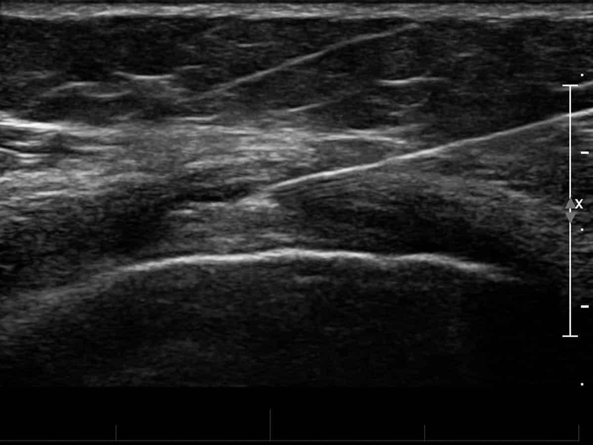
Ultrasound guided Hyaluronan injection into the medial aspect of the knee
Hyaluronan Injections
Hyaluronan is an injectable lubricant that may be effective in alleviating the symptoms of arthritis and is known by several trade names, such as Synvisc, Euflexxa, Supartz and Orthovisc. This procedure is known as viscosupplementation and replaces the lubricating properties of joint fluid that is depleted during arthritis. Hyaluronan results in improved joint cushioning and potentially provides a protective effect for joint cartilage cells. Symptomatic improvement has been demonstrated for up to 6 months, often delaying the need for joint replacement surgery. Hyaluronan may be injected into any joint affected by arthritis, most commonly the knee and hip.
Other conditions include:
- Chronic shoulder and hip pain
- Myofascial pain syndromes and trigger points
- Hamstring pain
- Groin pain, including osteitis pubis and adductor syndrome, including Femoral, Ilioinguinal and Genitofemoral nerve blocks
- Iliotibial band friction syndrome
- Sesamoiditis
- Prolotherapy
- Percutaneous trigger finger release
- Bone and soft tissue tumour biopsy.
Further Information.
Referring doctors are welcome to discuss with our radiologists the imaging and interventional radiology needs of their patients and whether a musculoskeletal injection is suitable for their patient’s medical condition.
Specialist Radiologists.